
Graphical Abstract
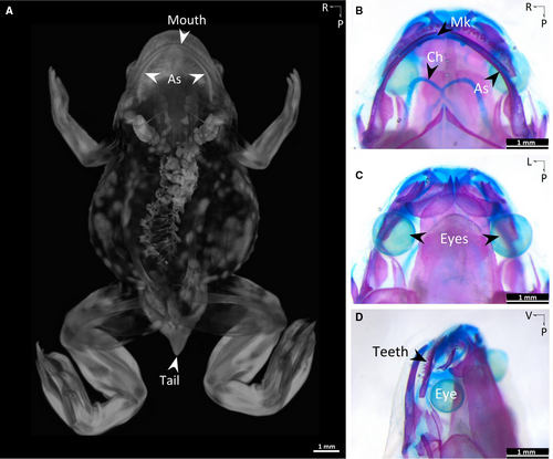
Dorsal view of a stage NF64 Xenopus tropicalis froglet stained with PTA (phosphotungstic acid) contrast agent, using inline phase-contrast imaging at the Canadian Light Source, the only synchrotron in Canada, demonstrates features of the nearlyadult frog skeleton, also showing soft tissues including visceral organs. The tail is nearly done resorbing as the froglet nears the completion of metamorphosis. The bulbous white structures on either side of the neck region are the otic capsules of the inner ear. This image was obtained in collaboration with SciArtist, Jean-Sébastien Gauthier (J-S Gauthier Creative Services). From: Common features of cartilage maturation are not conserved in an amphibian model; Jason K. B. Nguyen, Patsy Gómez-Picos, Yiwen Liu, Katie Ovens, B. Frank Eames. DevDyn 252:11; https://doi.org/10.1002/dvdy.594.
This 3D rendering illustrates the evolutionary repression of chondrogenic genes within vertebrate osteoblasts, as highlighted in the article ‘Evolutionary repression of chondrogenic genes in the vertebrate osteoblast’ published in the FEBS Journal (Issue 20, October 2020). The visualization showcases cellular structures and molecular details relevant to the research findings, emphasizing the intersection of scientific data and artistic representation.